


This article has been provided by Dr. Huseyin ACAR to give information about ‘Uveitis and Its Treatment’ in general terms.
HOW IS THE STRUCTURE OF OUR EYES ?
The main function of our eyes is to perceive the incoming light and to transmit it to the visual centers of our brain. In order to perform this function properly, three main layers made up the eye itself must be working in harmony. Sclera wraps around the eye like a shell to protect all the structures inside of the eye and is located at the outermost of our eyes. At the front side sclera becomes a transparent and called as cornea. There is the nerve layer called as retina located in the innermost of the eye. Uvea layer is placed between these two previous mentioned layers. Uveitis is the illness of this uvea layer. Uvea layer contains abundance of vessels and its main function is to feed the retina. Uvea layer consists of three sections: iris, ciliary body and choroid, in an order from front to the back.
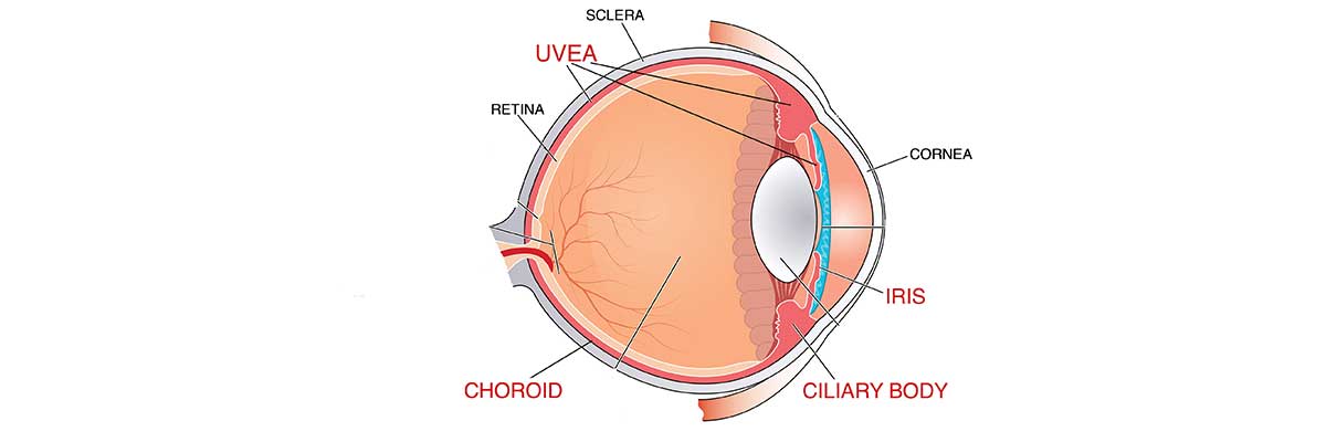
Image 1: Sclera (outermost), uvea (in the middle) and retina (innermost) are the layers of eye. Uvea can be subdivided into three sections; iris, ciliary body and choroid.
Uveitis is the inflammation of uvea layer of our eyes. Inflammation means the accumulation of defense cells in a certain specific area. Most frequent reason of inflammation is foreign organisms penetrating into our body. Another most frequent reason is the attack of our defense cells to our own tissues assuming as if they are foreign origin materials.
WHAT ARE THE SUB TYPES OF UVEITIS ?
We can separate the uvea layer from the front to the back by three sections: iris, ciliary body, and choroid. Inflammation may affect not only one of these sub sections but also more than one section. Taking all of this information into account, uveitis can be categorized into four sub groups. These are anterior uveitis, intermediate uveitis, posterior uveitis, and panuveitis where all uvea tissues are impacted.
WHAT ARE THE SYMPTOMS OF UVEITIS ?
Uveitis leads to different symptoms depending on the uveal area affected by the disease. If it affects the anterior part, then redness, pain and photophobia are the main complaints. If the middle area is affected, then the patient usually sees moving black objects. The main symptom is the reduction of vision in the posterior uveitis and panuveitis.
HOW TO DIAGNOS UVEITIS ?
Uveitis is generally diagnosed by an eye examination. In early stages, in case that clinical signs are not yet formed completely, then it would be difficult to make a diagnosis. After diagnosis, the determination of sub type or which sub section is impacted by disease must be done. Eye tests such eye angiography and OCT help us to determine which section is impacted. In addition these tests provide useful information about potential causes of the disease. Determination of the root cause forms mainstay of the treatment.
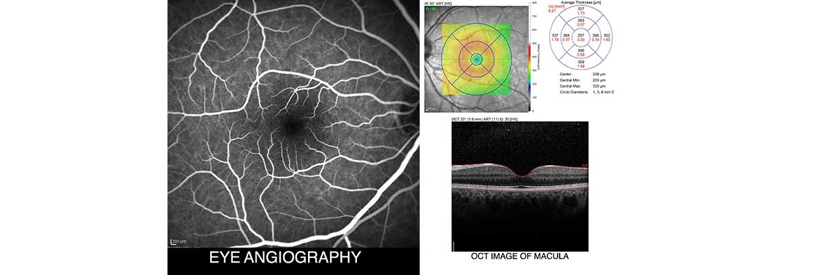
Image 2: Eye tests such eye angiography and OCT help us to determine which section is impacted. In addition these tests provide useful information about potential causes of the disease
HOW TO TREAT THE UVEITIS ?
Treatment is one of the most critical points in uveitis. Treatment is relatively easy if an underlying cause is found during examinations and tests. In case that the underlying cause is not found, the doctor will make a preliminary diagnosis according to his or her clinical experience and start treatment. Depending on the level of the response, he or she may make changes in the treatment over time. Another thing for treatment is the administration way of drugs. Eye drops is often sufficient in anterior uveitis. In case the inflammation is severe, injections into or around the eye may require. Eye drops alone are usually not enough for the types other than anterior uveitis. In the treatment of such uveitis, injections into or around the eye may be used alone or in combination with oral medicines.
SUMMARY
Uveitis is the inflammation of uvea layer of the eye. Inflammation may affect some or all of the uvea parts. Depending on the section affected by the disease, it shows itself with different symptoms. Diagnosis is usually made by an eye examination. Eye tests such as eye angiography and OCT help in confirming the diagnosis and investigating the underlying main cause. Treatment differs according to the affected area and the underlying problem.