


This article has been provided by Dr. Huseyin ACAR to give information about ‘Lacrimal Outflow System Obstruction in Adults and Its Treatment’ in general terms.
WHAT IS THE STRUCTURE OF LACRIMAL DUCT SYSTEM ?
Tear (or lacrima) is excreted by the lacrimal gland located at the upper part of our eyes. After it completes its function, it leaves the eye from the holes located at the upper and lower eyelids and then tear enters into the channel system. This channel system is mainly consisted of 6 parts: the first part is the holes. The holes exist at the upper and lower sections of the eyelids’ internal corners nearby the nose. They are not seen from outside but can be observed in case the eyelids are lifted towards outside. Lacrima enters into the channel system throughout these holes. The second part is the canaliculus. Lacrima is transferred into the lacrimal sac via these canaliculi. Afterwards it passes into the nasal cavity via lacrimal duct. It is only possible to discharge tears via these outflow system when our eyelids work properly. In each blink, our eyelids work like a pump and directs tears
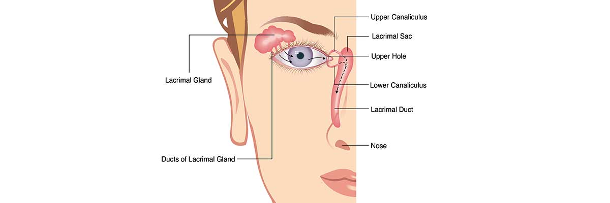
Image 1: Anatomy of tear outflow system
WHAT ARE THE SYMPTOMS OF LACRIMAL OUTFLOW SYSTEM OBSTRUCTION AND HOW TO DIAGNOSE IT?
Main complaint of the patients having lacrimal outflow system obstruction is the tearing. State of lacrimal outflow system for the people having the tearing problem is examined by means of various inspections and tests. Primarily under the microscope it is checked the position of lacrimal holes in the eyelids and whether they are obstructed or not. Then a thin cannula is inserted to the hole and liquid is administered. If the administered liquid falls easily into the throat of the patient, this means that system is not obstructed but if it does not, it means that the system is obstructed. There might also be some other tests required to be applied for some patient whom diagnosis cannot be done in a complete manner.
HOW TO TREAT LUCRIMAL OUTFLOW SYSTEM OBSTRUCTION IN ADULTS ?
Treatment of lacrimal outflow system obstruction in adults depends on the location of the obstruction. If obstruction is related to closure of the hole then the hole can be enlarged by a surgical operation or a silicon tube enabling liquid passage at the middle can be installed inside of the hole. This silicon tube may remain there for a long time and avoids any future obstruction. If obstruction occurs at a further point located beyond the lacrimal sac, this is the most of the cases, this time a new channel is required to be opened in bypassing the duct. This process is known as DSR operation. Today DSR operation has 3 techniques. The first of them is an external operation. This operation generally is done under general anesthesia and is realized by a skin cut over the lacrimal sac. Then a new channel is formed between lacrimal sac and nasal cavity. Sometimes a silicon tube may be inserted inside of this newly formed channel and that tube is removed after 3 to 6 months period. Advantage of such technique is that it has more than 90% success rate. This is the most successful technique when compared to other techniques.
The second technique is an endonasal DSR operation. This method is used together with inner nose camera or namely is done with accompanying endoscopy. New channel is formed between lacrimal sac and nasal cavity and a silicon tube is inserted into this channel. Advantage of this technique is that it has no skin cut. As for the disadvantage, it has less success rate than compared to the first technique. The third and the last technique is to open a channel between lacrimal sac and nasal cavity by the help of laser. After forming as large as possible channel, a silicon tube is inserted into this channel that will be removed after some time. Advantage of this technique is that it has no skin cut and disadvantage is that it has lower success ratio than two previous techniques.
SUMMARY
Lacrimal outflow system obstruction in adults leads to the complaint of tearing in patients. Obstruction usually occurs in the channel connecting the lacrimal sac and nasal cavity. In this case, a new channel between nasal cavity and lacrimal sac is required to be formed. There are many surgery techniques used for this purpose. The most successful technique with high ratio is the external method. There is no skin cut in the techniques realized inside of nose and using laser but these surgery methods have lower success rate than the first technique.