


This article has been provided by Dr. Huseyin ACAR to give information about ‘Retinal Vein Occlusion and Its Treatment’ in general terms.
WHAT IS THE STRUCTURE OF RETINA ?
Main task of our eyes is to perceive the incoming light and to transmit it to the visual centers of the brain. This is accomplished by focusing the incoming light upon the nerve layer, with the help of the lens system. The vessel system nourishing retina and removing the waste materials must work properly so that the retina can detect the incoming light and transmit it to the brain. The blood circulation in the retina consists of artery and vein systems. An artery passes through the optic nerve and reaches the retina and is divided into its branches. Then the system turns towards capillaries. Afterwards, the capillaries come together to form the veins. The veins are combined together to leave the eye through the optic nerve and carry the dirty blood to the heart.

Image 1: The blood circulation in the retina consists of artery and vein systems
WHAT ARE THE CAUSES OF RETINAL VEIN OCCLUSION ?
The main reason for occlusion in the retinal vein is the blood clots formed inside of the vessel. These clots are usually formed due to the deterioration of the vessel walls. Main reasons for the deterioration of vessel walls are the smoking, hypertension, cholesterol and diabetes. Additionally, high intraocular pressure may also cause occlusion of vein. Clots get formed in the vein with deteriorated inner wall structure caused for any reason. Clot does not prevent blood circulation and does not cause visual impairment unless it reaches to the certain level of size. After the size of the clot passes over the critical size, blood begins to accumulate in veins and capillaries. Intravenous blood and other liquids penetrate into the tissues of retina because of increased pressure inside of the vein and then damage the cells here.
WHAT ARE THE SYMPTOMS OF RETINAL VEIN OCCLUSIONS ?
Retinal vein occlusions must have reached above a certain level of occlusion in order to cause any symptoms. When such a condition is met, a painless reduction of vision occurs. Decrease in vision becomes even more pronounced if the occlusion level remains the same or increases. However, in case that our body is able to dissolve the clot, vision begins to improve slowly. The most important factor here is the level of occlusion of the veins and therefore the level of pressure within the vein.
HOW TO DIAGNOSE RETINAL VEIN OCCLUSION ?
In retinal vein occlusion, diagnosis is usually made with normal eye examination. Supporting tests such as eye angiography and OCT may be usually required in order to detect the level of disease and the impacted area. These tests are also used to follow the treatment response.
Şekil 2: Sağ gözümüzün anjiyosu. Hem atardamarlar, hem de toplardamarlar damar içinden verilen florosein maddesi ile dolduğu için beyaz renkte görünmektedir. Sıvı kaçağı olmadığı için damarların dışındaki bölgeler siyah renklidir.
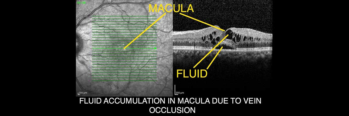
Image 2: A patient with retinal vein occlusion. There is fluid accumulation between retinal layers
HOW TO TREAT RETINAL VEIN OCCLUSION ?
Treatment type of retinal vein occlusion is decided according to the results of the examination and the applied tests. Treatment is administered in case that the vision has decreased or would be decreased if not treated. Methods used in treatment are intraocular injections and argon laser. Generally intraocular injections are applied when the vision is decreased due to the accumulation of liquid at macula (centre of retina). As for laser treatment, it is mostly used when retinal tissue located outer side of macula is severely damaged. Laser treatment usually does not contribute to the increase in vision. It simply avoids new vessel formation which may cause bleeding in the eye. The whole purpose here is to preserve existing vision and to prevent some undesired situations that may be prevailed in future.
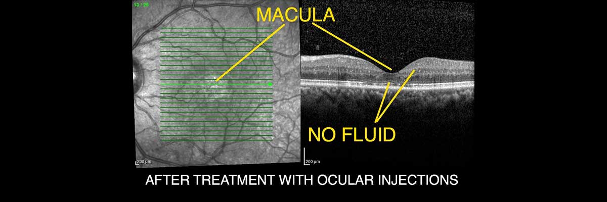
Image 3: Same patient in image 2. Fluid disappeared after treatment with intraocular injections and vision increased
The reason of retinal vein occlusion is generally the clots that are formed due to the deterioration of vein walls. Most of the time, the formed clots are cleaned by the body. In case that they were not able to be removed by the body, it cause obstruction and accumulation of blood in certain places and leads to harm of nerve cells. Retinal vein occlusion manifests itself as visual disturbance. Intraocular injections and argon laser are used for the treatment of disease. It is decided according to results of eye examination and tests results.